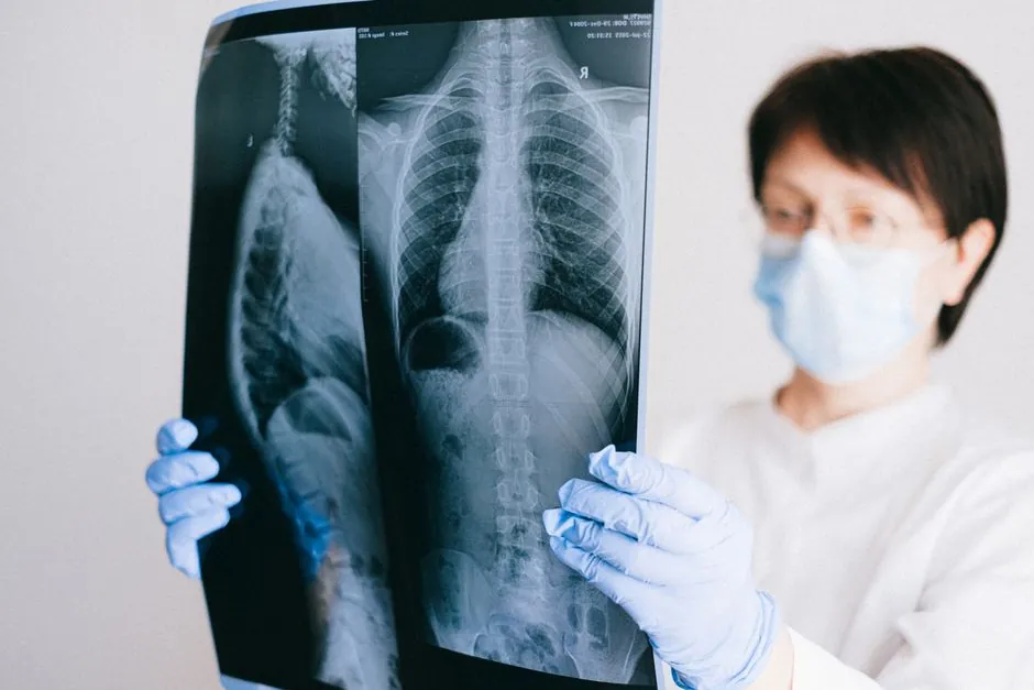
Flat-panel detectors (FPD) and image intensifiers (II) sit on opposite ends of a real-time radioscopy decision, and the gap is technical, not cosmetic: FPD removes the optical conversion step entirely, while II still runs X-ray → light → electron → light → camera [S3].
The core spec trade is resolution and dose stability versus capital cost and a smaller room footprint. FPD uses a scintillator (CsI or Gd2O2S) coupled to an amorphous-silicon photodiode array and outputs digital data directly; II amplifies light through a vacuum tube, then re-images it through a TV camera [S2][S3].
How the two detector chains actually differ
II/TV has been the workhorse of fluoroscopy for over 60 years, with the X-ray image intensifier introduced in the late 1940s and optically coupled to a TV camera for closed-circuit display [S1]. In an II, X-rays strike an input phosphor, are converted to electrons at a photocathode, accelerated and minified through a vacuum tube, and re-emitted at an output phosphor, where a camera digitises the brightened image [S3][S6]. Each optical-electronic conversion adds noise, and the convex input phosphor forces peripheral geometric distortion and veiling glare into the chain [S1][S3].
FPD eliminates that chain. An FPD is built around a scintillator layer (CsI or Gd2O2S) bonded to an amorphous-silicon (a-Si) photodetector array; the scintillator absorbs X-rays, the photodiode array turns the resulting light into charge, and the signal is digitised on the panel itself with no optical relay, no minification, and no TV bandwidth limit [S2][S6]. Two implementation paths exist: indirect-conversion (scintillator + photodiode) and direct-conversion (semiconductor absorbing X-ray photons directly), and both are widely used in modern interventional fluoroscopy [S1].
Resolution, distortion, and magnification behaviour
Resolution and distortion are where FPD wins by construction. The flat input surface and pixel-addressable readout remove the geometric (pincushion) distortion inherent in an II's curved input phosphor, and FPDs show minimal image degradation across years of service versus the gradual gain loss seen on image intensifiers [S3][S4]. A direct comparison often quoted by OEMs: "FPDs offer more consistent image quality and a larger field of view, while image intensifiers are often more cost-effective and provide strong magnification capabilities" [S5].
Magnification is a clean spec story on both sides, but the dose consequence differs. With an II, magnification is achieved by electronic collimation, which crops the field of view, and dose must be increased to keep the magnified image bright; with an FPD, the same digital zoom is achieved with no increase in radiation to the patient [S3][S4]. For dose-sensitive paediatric interventional work, where real-time fluoroscopy is becoming more prevalent, this difference is the central selection driver [S1].
Dose, detective quantum efficiency, and ALARA
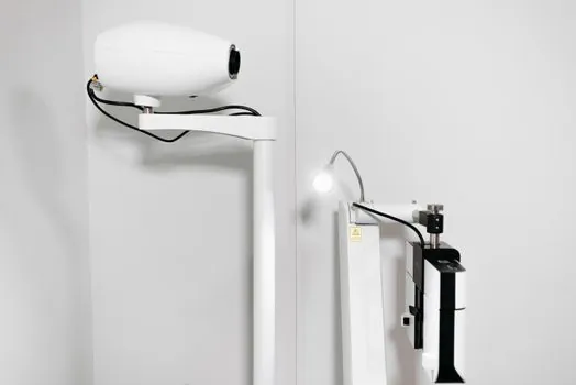
Multiple sources state, in nearly identical wording, that FPD systems use less radiation than image intensifiers for the same diagnostic task, and that FPDs are "more sensitive compared to image intensifiers," which "lower[s] the patient dose" [S3][S4][S7]. The underlying mechanism is better detective quantum efficiency (DQE) on the FPD chain, which makes it possible to reduce exposure "in accordance with ALARA principles" [S1].
The historical caveat still matters: at the lowest fluoroscopic exposure levels, first-generation FPDs could not match II/TV image quality, and they showed residual signal from previous exposures and limited tableside protocol control [S1]. Second-generation and hybrid FPDs have largely closed that gap, which is why the technology has spread from interventional suites into mini C-arms and surgical C-arms [S1][S5][S8].
Form factor, ergonomics, and footprint
II is a long vacuum tube on a C-arm, while FPD is a flat panel mounted at the detector end. The flat-panel detector is "more compact when compared to the extended tube structure of an image intensifier," giving the operator more working space and better ergonomics around larger patients [S4]. Mini C-arms with image intensifiers, such as the Orthoscan HD and Hologic Fluoroscan InSight 2, remain in production precisely because the smaller II tube preserves a tight arc depth and vertical range for extremity work [S5].
Field of view also differs at the same panel size. II systems lose active area as the input magnification mode narrows the beam; FPDs hold usable pixels across the full square or rectangular active area, which is why OEM literature keeps calling out the "larger field of view" of FPD chains [S5]. For positioning the detector close to the patient, a compact FPD head also reduces source-to-image distance (SID) loss on portable and mini platforms.
Cost, service life, and total cost of ownership
Upfront capital, FPD costs more. Refurbished image intensifier C-arms are widely available in the secondary market and span a wide price band; refurbished FPD systems are scarcer and command a premium, because the panel itself is the single most expensive sub-assembly [S3]. For new build-outs, this is partly offset over the asset's life, because FPD image quality does not degrade the way an II's does, and long-term II tube replacement plus higher repair frequency raise the total cost of ownership on the older platform [S4].
For a low-to-moderate patient volume facility, or one whose C-arm use is limited to general surgery and standard fluoroscopic cases, an image intensifier is often the right call, and that is how several vendors frame the decision in their own product copy [S3][S5]. For high-volume interventional, vascular, or paediatric work, the FPD cost premium typically pays back through dose, uptime, and image-consistency gains [S3][S4].
Selection matrix: when to pick which
Pick FPD when: dose reduction is mandated (paediatric, interventional, ALARA-driven programmes), image consistency over 5-10 years matters, you need a square large FOV without dose penalty on zoom, or you have a fixed suite and want compact ergonomics [S1][S3][S4]. Pick II when: capital budget is tight, a refurbished platform meets the use case, the room is small and the long II tube is not a constraint, or patient volume is low-to-moderate and general surgery is the main application [S3][S5][S7]. If your facility is more analogous to a moving assembly cell than a fixed line, the maintenance access story also matters; FPD panels are sealed units that swap out cleanly, while II tubes need periodic gain and focus calibration.
Cross-reference the engineering practice of mapping detector choice to procedure class, not to brand. A standard linear decision rule that vendors apply is: orthopaedic extremity and pain-management work on mini C-arms stays on II, while vascular, cardiac, and complex interventional migrates to FPD [S5][S8]. Sites that bridge both worlds often standardise on FPD to remove a parallel detector fleet, accepting higher capex for simpler training and one QA workflow [S4][S8].
What the data still cannot tell you
Several claims in the public sources are repeated but not numerically backed. Statements like "FPD shows minimal image degradation over a longer period" or "II produces lower resolution" are universally agreed across vendors, but no public source in the research set gives a quantified DQE figure, a contrast-detail curve, or a MTF in lp/mm for either detector class at a given kVp [S3][S4][S5]. For procurement, that means the "FPD is better" line is directionally correct but not benchmarked here in numbers; any site-specific decision should pull the OEM's measured DQE at the operating kVp, the stated pixel pitch (typically 100-200 µm for fluoroscopy FPDs), and the frame rate (commonly up to 30 fps in real-time fluoroscopy, higher in pulsed modes) before signing the spec.
Related specification disciplines that share the same decision logic (capex vs. lifetime performance, digital vs. legacy analog chain) are covered elsewhere on the site: see forged vs rolled alloy steel bar selection for material selection under cyclic loading, VLM vs carousel for footprint-vs-throughput trades, and S/FTP vs F/UTP vs U/FTP for the equivalent analog-vs-digital decision in cabling. Detector choice in fluoroscopy follows the same pattern: the newer digital chain wins on every measurable performance axis, and the legacy chain survives only where capex and footprint dominate.
For the relevant spec sheets and selection criteria, see flat belt, time relay, and alc panel.